38 external structure of the heart with labels
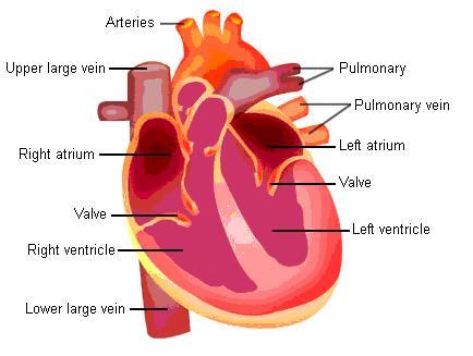
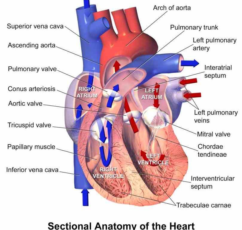
Human Heart (Anatomy): Diagram, Function, Chambers, Location in Body The heart is a muscular organ about the size of a fist, located just behind and slightly left of the breastbone. The heart pumps blood through the network of arteries and veins called the ... A Guide on the Structure of heart, its Chambers and valves Internal anatomy of the Heart. The heart is divided into the right and left side due to a septum which is made of myocardium and endocardium. Each side is divided into the upper atrium and lower ventricle due to atrioventricular valves. Thus, there are four chambers in a human heart. Each right and left halves contain one atrium and one ...
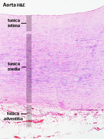
Layers of the heart: Epicardium, myocardium, endocardium | Kenhub The myocardium is functionally the main constituent of the heart and the thickest layer of all three heart layers. It is a muscle layer that enables heart contractions. Histologically, the myocardium is comprised of cardiomyocytes.Cardiomyocytes have a single nucleus in the center of the cell, which helps to distinguish them from skeletal muscle cells that have multiple nuclei dispersed in the ...

External structure of the heart with labels
The 3 Layers of the Heart Wall - ThoughtCo The heart is an extraordinary organ. It is about the size of a clenched fist, weighs about 10.5 ounces and is shaped like a cone. Along with the circulatory system, the heart works to supply blood and oxygen to all parts of the body. The heart is located in the chest cavity just posterior to the breastbone, between the lungs, and superior to the diaphragm. Heart Anatomy Labeling Game This is an online quiz called Heart Anatomy Labeling Game. There is a printable worksheet available for download here so you can take the quiz with pen and paper. Your Skills & Rank. Total Points. 0. Get started! Today's Rank--0. Today 's Points. One of us! Game Points. 19. You need to get 100% to score the 19 points available. External anterior heart labeling Quiz - PurposeGames.com This is an online quiz called External anterior heart labeling There is a printable worksheet available for download here so you can take the quiz with pen and paper. Your Skills & Rank Total Points 0 Get started! Today's Rank -- 0 Today 's Points One of us! Game Points 27 You need to get 100% to score the 27 points available Actions
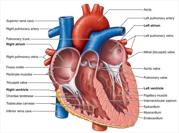
External structure of the heart with labels. byjus.com › biology › diagram-of-heartHeart Diagram with Labels and Detailed Explanation - BYJUS Diagram of Heart. The human heart is the most crucial organ of the human body. It pumps blood from the heart to different parts of the body and back to the heart. The most common heart attack symptoms or warning signs are chest pain, breathlessness, nausea, sweating etc. The diagram of heart is beneficial for Class 10 and 12 and is frequently ... The Anatomy of the Heart, Its Structures, and Functions Updated on April 05, 2020. The heart is the organ that helps supply blood and oxygen to all parts of the body. It is divided by a partition (or septum) into two halves. The halves are, in turn, divided into four chambers. The heart is situated within the chest cavity and surrounded by a fluid-filled sac called the pericardium. Ch. 19 Circulatory System- heart Flashcards | Quizlet Correctly label the external anatomy of the anterior heart. Place the labels in order denoting the flow of blood through the pulmonary circuit beginning with the right atrium and ending in the left atrioventricular valve. The first and last structures are given. Right atrium 1. tricuspid valve 2. right ventricle 3. pulmonary valve Internal Structure of the Heart | Contemporary Health Issues It is marked by the presence of four openings that allow blood to move from the atria into the ventricles and from the ventricles into the pulmonary trunk and aorta. Located in each of these openings between the atria and ventricles is a valve, a specialized structure that ensures one-way flow of blood.
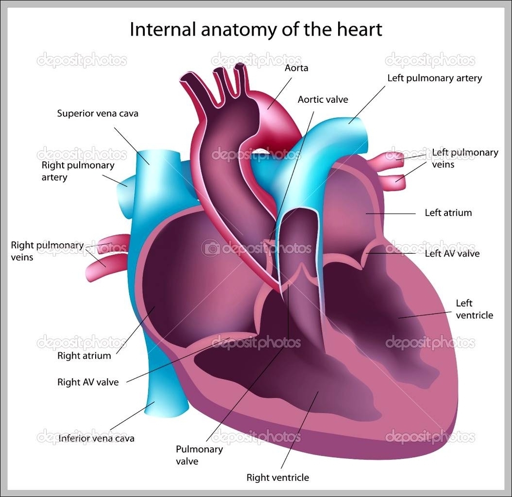
How to Draw the Internal Structure of the Heart (with Pictures) To draw the internal structure of a human heart, follow the steps below. Part 1 Finding a Diagram 1 To find a good diagram, go to Google Images, and type in "The Internal Structure of the Human Heart". Find an image that displays the entire heart, and click on it to enlarge it. 2 Find a piece of paper and something to draw with. Heart Anatomy: size, location, coverings and layers - Anatomy & Physiology The heart wall is composed of three layers: the epicardium, myocardium, and endocardium. Location of the heart in the mediastinum. The superficial epicardium is the visceral layer of the serous pericardium. The middle layer is the myocardium and is composed mainly of cardiac muscle and forms the bulk of the heart. › heart › structure-heartStructure of the Heart | The Franklin Institute Structure of the Heart Although most people know that the human heart doesn’t bear much resemblance to a heart drawn on a Valentine’s Day card, the image can still be a useful way to learn and remember the parts of the heart. The heart consists of four chambers: two atria on the top and two ventricles on the bottom. Human Heart - Anatomy, Functions and Facts about Heart The heart wall is made up of 3 layers, namely: Epicardium - Epicardium is the outermost layer of the heart. It is composed of a thin-layered membrane that serves to lubricate and protect the outer section. Myocardium - This is a layer of muscle tissue and it constitutes the middle layer wall of the heart.
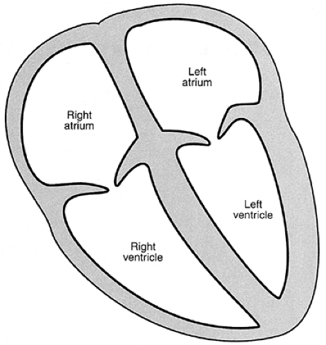
Chapter 22 Heart Flashcards | Quizlet Label the coronary arteries in an anterior view of the heart. Label the order that blood flows through in the heart, using the arrows as guides. Label the components of the heart wall. Label the components of the heart as seen from a posterior view. Label the major coronary veins. Label the components of the conduction system. Diagram of the human heart Images, Stock Photos & Vectors - Shutterstock Find Diagram of the human heart stock images in HD and millions of other royalty-free stock photos, illustrations and vectors in the Shutterstock collection. Thousands of new, high-quality pictures added every day. ... Anatomy. Healthcare and Medical. Diseases, Viruses, and Disorders. Icons and Graphics. heart. medicine. organ. human body. Structure Of The Heart | A-Level Biology Revision Notes The heart is a hollow muscular organ that lies in the middle of the chest cavity. It is enclosed in the pericardium, which protects the heart and facilitates its pumping action. The heart is divided into four chambers: The two atria (auricles): these are the upper two chambers. They have thin walls which receive blood from veins. The structure of the heart - Structure and function of the heart ... Each side of the heart consists of an atrium and a ventricle which are two connected chambers. The atria (plural of atrium) are where the blood collects when it enters the heart. The ventricles...
Know the Structures and Functions about Your Heart | New Health Advisor
Label the Heart - The Biology Corner Shows a picture of a heart with letters and blanks for practice with labeling the parts of the heart and tracing the flow of blood within the heart.

CLASS BLOG: BIO 202 Heart Anatomy Worksheet
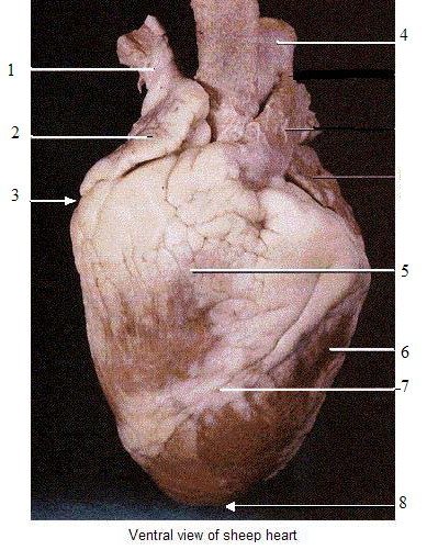
Heart Anatomy: Heart Dissection The letters indicated in the text refer to the labels on the picture. The anterior surface of the heart is characterized by the presence of the large arteries leaving the base of the heart, the pulmonary trunk (H) and the aorta (G). The pulmonary trunk is the vessel that divides to give rise to the two pulmonary arteries going to each lung.
ANAT2241 Cardiovascular System - Embryology
› 1-label-the-heartLabel the heart — Science Learning Hub Jun 16, 2017 · In this interactive, you can label parts of the human heart. Drag and drop the text labels onto the boxes next to the diagram. Selecting or hovering over a box will highlight each area in the diagram. Pulmonary vein Right atrium Semilunar valve Left ventricle Vena cava Right ventricle Pulmonary artery Aorta Left atrium Download Exercise Tweet

fantasticvoyagewiki / Heart
Solved Art-Labeling Activity: Overview of the external - Chegg art-labeling activity: overview of the external anatomy of the heart anterior view res great cardiac vein aortic arch right coronary artery left coronary artery left pulmonary veins ascending aorta left pulmonary artery anterior interventricular artery superior vena cava pulmonary trunk auricle of left atrium circumflex artery auricle of right …

The Cardiovascular System: Anatomy & Physiology - The Nursing Journal
bodytomy.com › labeled-diagram-of-human-heartA Labeled Diagram of the Human Heart You Really Need to See The human heart, comprises four chambers: right atrium, left atrium, right ventricle and left ventricle. The two upper chambers are called the left and the right atria, and the two lower chambers are known as the left and the right ventricles. The two atria and ventricles are separated from each other by a muscle wall called ‘septum’.
13+ Heart Diagram Templates – Sample, Example, Format Download | Free & Premium Templates
Surface projections of the heart: Borders and landmarks - Kenhub 1/4. The surface projections of the heart represent points on the thoracic wall that map out the outline and valves of the heart. These include four borders (superior, right, inferior, left) and four valves (left atrioventricular, right atrioventricular, aortic, pulmonary). The main reference points used for the surface projections of the heart ...

Lung Structure | BioNinja
Heart - External Features - Anatomy QA Location of heart: Heart lies in the middle mediastinum. 1/3rd of the heart lies to the right and 2/3rd to the left of the midline. It lies opposite to T5 - T8 vertebrae in supine position & T6 - T9 vertebrae in erect position. Dimensions of heart: Base to apex-12cm; Transversely- 8-9cm; Anteroposteriorly- 6cm.
Structure and Function of the Normal Heart | Thoracic Key
OpenStax AnatPhys fig.19.6 - Surface Anatomy of the Heart - English labels External Anatomy of the Heart. Inside the pericardium, the surface features of the heart are visible. English labels. From OpenStax book 'Anatomy and Physiology', fig. 19.6. Anatomical structures in item: Cor Truncus brachiocephalicus Vena cava superior Truncus pulmonalis Aorta ascendens Atrium dextrum Atrium sinistrum Ventriculus dexter
called myocardium science External Structure Of Human Heart Anatomy structure of human heart ...
Solved Label the external anatomy on this anterior view of a | Chegg.com Label the external anatomy on this anterior view of a mammalian heart by clicking and dragging the labels to the correct location 8 Apex of heart Pulmonary trunk Right ventricle Right coronary artery (RCA) Right marginal branch of RCA Right uncle Left ventricle Left pulmonary artery Antenor interventricular branch of LGA Ascending aorta Zoom Reset

Things cardiologists never tell you: KNOW YOUR HEART. Structure of the heart and blood vessels ...
Heart Anatomy | Anatomy and Physiology - Lumen Learning The wall of the heart is composed of three layers of unequal thickness. From superficial to deep, these are the epicardium, the myocardium, and the endocardium. The outermost layer of the wall of the heart is also the innermost layer of the pericardium, the epicardium, or the visceral pericardium discussed earlier. Figure 6.
37 Label The Anatomy Of The Heart - Labels 2021
encounteredu.com › teacher-resources › googleLesson | The Heart - External Structure | Encounter Edu In this lesson students begin their exploration of the circulatory system, labelling a diagram of the external structures and identifying arteries and veins. They will go on to explain where blood enters and leaves the heart. Learning outcomes

Labels Of The Heart Diagram - sharedoc
Human Heart - Diagram and Anatomy of the Heart - Innerbody Because the heart points to the left, about 2/3 of the heart's mass is found on the left side of the body and the other 1/3 is on the right. Anatomy of the Heart Pericardium. The heart sits within a fluid-filled cavity called the pericardial cavity. The walls and lining of the pericardial cavity are a special membrane known as the pericardium.

what is heart!?what is it function? and structure - Brainly.in
External anterior heart labeling Quiz - PurposeGames.com This is an online quiz called External anterior heart labeling There is a printable worksheet available for download here so you can take the quiz with pen and paper. Your Skills & Rank Total Points 0 Get started! Today's Rank -- 0 Today 's Points One of us! Game Points 27 You need to get 100% to score the 27 points available Actions

Heart Anatomy Labeling Game This is an online quiz called Heart Anatomy Labeling Game. There is a printable worksheet available for download here so you can take the quiz with pen and paper. Your Skills & Rank. Total Points. 0. Get started! Today's Rank--0. Today 's Points. One of us! Game Points. 19. You need to get 100% to score the 19 points available.

Free Blank Heart Diagram, Download Free Blank Heart Diagram png images, Free ClipArts on Clipart ...
The 3 Layers of the Heart Wall - ThoughtCo The heart is an extraordinary organ. It is about the size of a clenched fist, weighs about 10.5 ounces and is shaped like a cone. Along with the circulatory system, the heart works to supply blood and oxygen to all parts of the body. The heart is located in the chest cavity just posterior to the breastbone, between the lungs, and superior to the diaphragm.
Heart Model Contiunued - ProProfs Quiz

Heart Diagram
Post a Comment for "38 external structure of the heart with labels"