40 simple microscope diagram with labels
pages.zeiss.com › rs › 896-XMS-794Principles of Fluorescence and Fluorescence Microscopy - ZEISS The Fluorescence Microscope The main requirement of a fluorescence microscope is to illuminate a specimen with light of an excitatory wavelength whilst simultaneously collecting and separating the compara-tively weaker light emitted by the sample. In the example of Stokes’ observation, these tasks are performed by the Microscope Drawing And Label at PaintingValley.com ... label microscope diagram compound parts light labeling functions microscopic blank labeled biology microscopy labelled beautiful Compound Microscope ... 496x600 35 0 Parts Of A Compound ... 500x469 27 0 Microscopic Drawing ... 1024x1024 21 4 Download The Diagram... 547x579 17 0 Microscope Labeling ... 270x350 17 0 Microscope Labeling ...
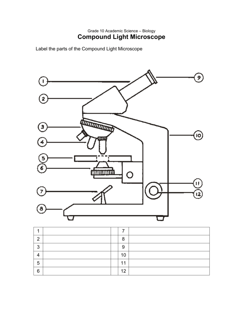
Parts of the Microscope with Labeling (also Free Printouts ... Parts of the Microscope with Labeling (also Free Printouts) A microscope is one of the invaluable tools in the laboratory setting. It is used to observe things that cannot be seen by the naked eye. Table of Contents 1. Eyepiece 2. Body tube/Head 3. Turret/Nose piece 4. Objective lenses 5. Knobs (fine and coarse) 6. Stage and stage clips 7. Aperture

Simple microscope diagram with labels
quizlet.com › 290507793 › chapter-10-masteringbioChapter 10 MasteringBio Homework Flashcards - Quizlet Drag the labels onto the flowchart to show the relationship between the production of photons by the sun (Engelmann's light source) and the distribution of bacteria that Engelmann observed under his microscope. Not all labels will be used. Compound Microscope- Definition, Labeled Diagram ... The optical microscope often referred to as the light microscope, is a type of microscope that uses visible light and a system of lenses to magnify images of small subjects. There are two basic types of optical microscopes: Simple microscopes. Compound microscopes. The term "compound" in compound microscopes refers to the microscope having ... Compound Microscope Parts - Labeled Diagram and their ... There are three major structural parts of a compound microscope. The head includes the upper part of the microscope, which houses the most critical optical components, and the eyepiece tube of the microscope. The base acts as the foundation of microscopes and houses the illuminator. The arm connects between the base and the head parts.
Simple microscope diagram with labels. PDF Parts of a Microscope Printables - Homeschool Creations Label the parts of the microscope. You can use the word bank below to fill in the blanks or cut and paste the words at the bottom. Microscope Created by Jolanthe @ HomeschoolCreations.net eyepiece head objective lenses arm focusing knob base illuminator stage stage clips nosepiece. Label Microscope Diagram - EnchantedLearning.com arm - this attaches the eyepiece and body tube to the base. base - this supports the microscope. body tube - the tube that supports the eyepiece. coarse focus adjustment - a knob that makes large adjustments to the focus. diaphragm - an adjustable opening under the stage, allowing different amounts of light onto the stage. Simple Microscope Definition, Magnification, Parts And Uses Aim: To make a simple microscope with the help of water. Apparatus Required A glass of water Fuse wire Object to view (newspaper works well due to its fine print) Procedure Make a loop of the fuse wire around 2 mm wide. Dip it in water so that a drop is made in the loop. Hold it near to your eye and take a close look at the object you have chosen. Simple Columnar Epithelium Labeled Diagram Squamous means scale-like. simple squamous. Bodytomy provides a labeled diagram to help you understand the structure and Simple Columnar Epithelium: Labeled Diagram and Function. Epithelium is a tissue that lines the internal surface of the body, as well as the internal organs. Simple epithelium is one of the types of epithelium that is.
Label the Microscope Diagram | Download Scientific Diagram the antibiogram of e. coli was investigated in different generations using eight antibiotic discs such as chloramphenicol (ch), streptomycin (s), gentamycin (g), ciprofloxacin (ci),... Compound Microscope Parts, Functions, and Labeled Diagram ... Compound Microscope Definitions for Labels. Eyepiece (ocular lens) with or without Pointer: The part that is looked through at the top of the compound microscope. Eyepieces typically have a magnification between 5x & 30x. Monocular or Binocular Head: Structural support that holds & connects the eyepieces to the objective lenses. Parts of a Simple Microscope - Labeled (with diagrams ... Parts of a Simple Microscope - Labeled (with diagrams) A simple microscope is a very first type of microscope ever created. It consists of simple parts and performs simple functions. Although there are now many advanced microscope types, some applications may still demand the use of a simple microscope. Simple Microscope - Parts, Functions, Diagram and ... Simple Microscope - Parts, Functions, Diagram and Labelling A microscope is one of the commonly used equipment in a laboratory setting. A microscope is an optical instrument used to magnify an image of a tiny object; objects that are not visible to the human eyes. Table of Contents The common types of microscopes are: What is a Simple microscope?
Microscope Poster - Diagram with Labels | Teach Starter A poster containing a diagram with labels showing the key parts of a microscope. In Science it is important that students know how to use a variety of tools when conducting scientific experiments and inquiry. This poster focuses on the microscope and highlights its key parts. There are two print options available for this poster: A Study of the Microscope and its Functions With a Labeled ... sciencestruck.com A Study of the Microscope and its Functions With a Labeled Diagram To better understand the structure and function of a microscope, we need to take a look at the labeled microscope diagrams of the compound and electron microscope. These diagrams clearly explain the functioning of the microscopes along with their respective parts. Types of Microscopes: Definition, Working Principle ... Where, D is the least distinct vision; F is the focal length of the convex lens; Simple Microscope Diagram. Principle of Simple Microscope. The working principle of a simple microscope is that when a sample is placed within the focus of the microscope, a virtual, erect and magnified image is obtained at the least distance of distinct vision from the eye that is held at the lens. Parts of a microscope with functions and labeled diagram Figure: Diagram of parts of a microscope There are three structural parts of the microscope i.e. head, base, and arm. Head - This is also known as the body. It carries the optical parts in the upper part of the microscope. Base - It acts as microscopes support. It also carries microscopic illuminators.
All Saints Online
Microscope, Microscope Parts, Labeled Diagram, and Functions Microscope, Microscope Parts, Labeled Diagram, and Functions What is Microscope? A microscope is a laboratory instrument used to examine objects that are too small to be seen by the naked eye. It is derived from Ancient Greek words and composed of mikrós, "small" and skopeîn,"to look" or "see".

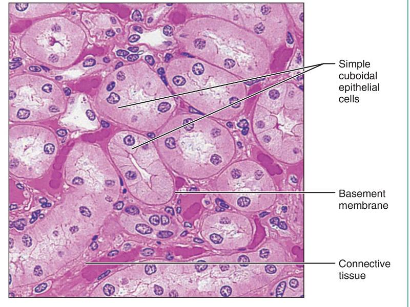
31 best images about Histology - GI - Layers, Junctions and Miscellaneous on Pinterest
Microscope Labeling - The Biology Corner 1) Start with scanning (the shortest objective) and only use the COARSE knob . Once it is focused… 2) Switch to low power (medium) and only use the COARSE knob . You may need to recenter your slide. Once it is focused.. 3) Switch to high power (long objective).

The Wonderful Microworld: Cell Nucleus - Onion
quizlet.com › 568033559 › botany-exam-1-chs-1/2/3-4Botany Exam 1 Chs. 1, 2, 3, 4, 5, 6, 7, 12, and 16 Quizzes Mitosis, or the division of a mother cell's nucleus into two identical daughter nuclei, is typically divided into four phases. Match each of the labels to identify what events take place during each phase of mitosis. 1. Prophase 2. Metaphase 3. Anaphase 4. Telophase A) Chromosomes are aligned at the equator of the cell and the spindle is fully ...
33 Microscope Diagram To Label - Labels Database 2020
Simple Microscope - Definition, Diagram, FAQs Simple Microscope Definition: A Simple Microscope meaning is used to see a magnified image of an object. Antonie Van Leeuwenhoek, a Dutchman, invented the first simple microscope, consisting of a single powerful magnetic lens that rotates to detect tiny freshwater insects. It is composed mainly of light microscopes.

www.timvandevall.com wp-content uploads Labeled-Microscope-Diagram.jpg | ชีววิทยาศาสตร์, ห้อง ...
Label a Microscope - Storyboard That Create a poster that labels the parts of a microscope and includes descriptions of what each part does. Click "Start Assignment". Use a landscape poster layout (large or small). Search for a diagram of a microscope. Using arrows and textables label each part of the microscope and describe its function. Copy This Storyboard* More options
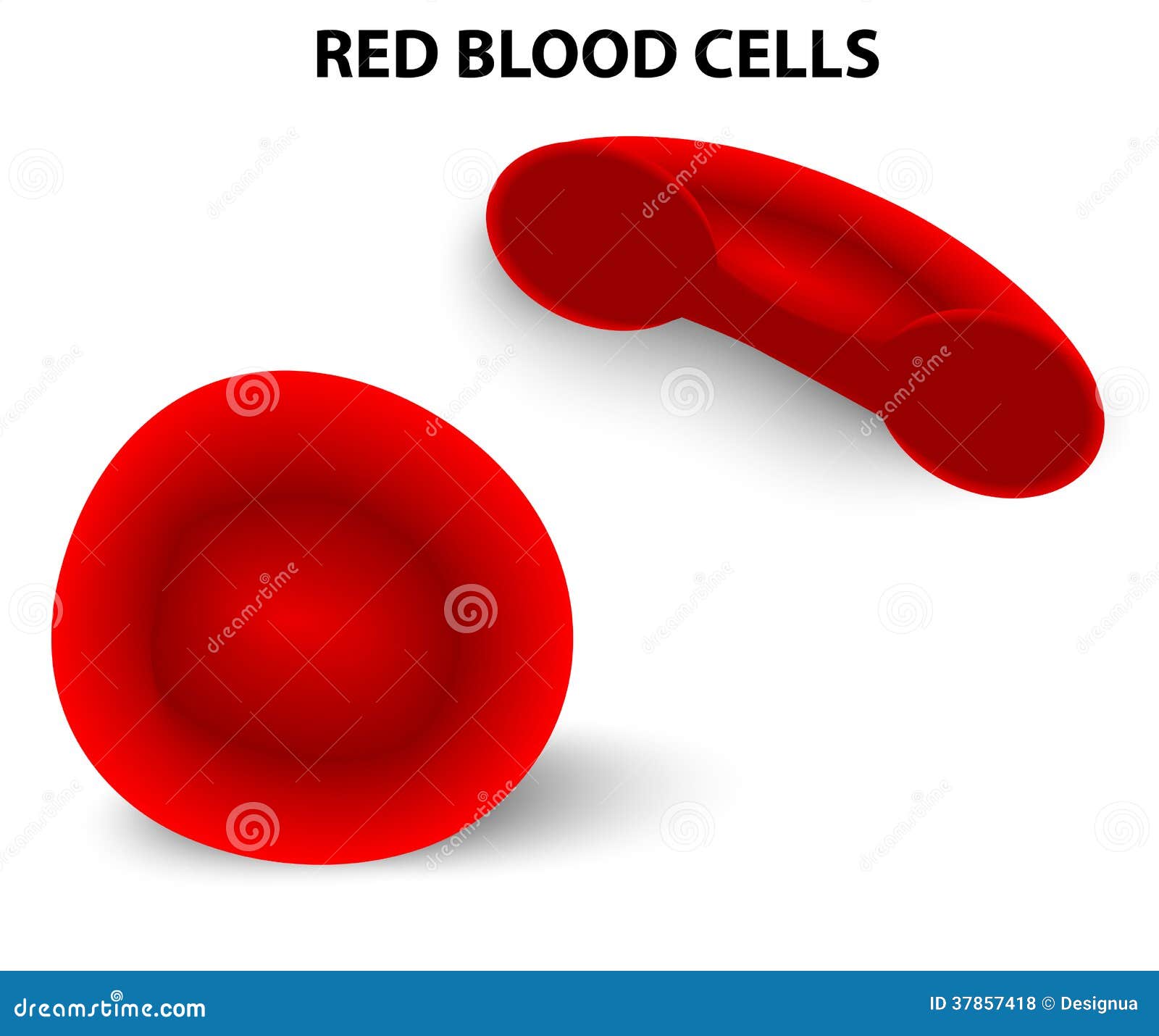
Red blood cells stock vector. Image of blood, healthy - 37857418
Microscope Parts and Functions With Labeled Diagram and ... Microscope Parts and Functions With Labeled Diagram and Functions How does a Compound Microscope Work?. Before exploring microscope parts and functions, you should probably understand that the compound light microscope is more complicated than just a microscope with more than one lens.. First, the purpose of a microscope is to magnify a small object or to magnify the fine details of a larger ...

microscope labeled microscope worksheet labeling sc 1 st template entrancing labelling - Top ...
Compound Microscope Parts - Labeled Diagram and their ... There are three major structural parts of a compound microscope. The head includes the upper part of the microscope, which houses the most critical optical components, and the eyepiece tube of the microscope. The base acts as the foundation of microscopes and houses the illuminator. The arm connects between the base and the head parts.

Microscope Diagram Unlabeled - Micropedia
Compound Microscope- Definition, Labeled Diagram ... The optical microscope often referred to as the light microscope, is a type of microscope that uses visible light and a system of lenses to magnify images of small subjects. There are two basic types of optical microscopes: Simple microscopes. Compound microscopes. The term "compound" in compound microscopes refers to the microscope having ...

Amoeba Cell Diagram
quizlet.com › 290507793 › chapter-10-masteringbioChapter 10 MasteringBio Homework Flashcards - Quizlet Drag the labels onto the flowchart to show the relationship between the production of photons by the sun (Engelmann's light source) and the distribution of bacteria that Engelmann observed under his microscope. Not all labels will be used.

Q14 Draw a large diagram of an animal cell as seen through an electron microscope. Label the ...

Label Microscope Diagram - ClipArt Best
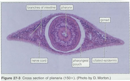
Flatworms
Print Anatomy Midterm 2 flashcards | Easy Notecards

Porifera diagram | Science biology, Arthropods, Plant science
Post a Comment for "40 simple microscope diagram with labels"